Cell : The Unit of Life Selina biology Class 9 chapter 2
Cell is the structural and functional unit of all living organism all living organism are made up of cell
Cell : The Unit of Life Selina biology Class 9 chapter 2
Cell –
It is the Fundamental Structural and Functional Unit of the all living organism

https://en.wikipedia.org/wiki/Cell_(biology)
All living organism are made up of the cell
The Invention of the Microscope –

ANTON VAN LEEUWENHOEK –
- He was a Dutch Scientist
- A. V. Leeuwenhoek constructed 1st Microscope
- he made many microscope
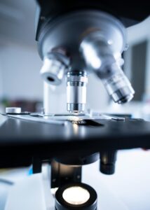
- the microscope made by Leeuwenhoek are known as ” Simple Microscope”
- They are called as Simple microscope because it consist of ” a Single Biconvex Lens”
Cell : The Unit of Life Selina biology Class 9 chapter 2
Discovery of cell –
Robert Hook ( 18 July 1635 – 3 March 1703) –
- He Constructed Compound Microscope
- it is known as Compound Microscope because it was developed by using Two Lenses
- he observed ” Slice of cork” and find that it was made up of ” Box/ Compartment like Structure “
- he gives it name ” Cell” after Latin name ” Cella” meaning ” Small Room”
Microbes in Human Welfare NCERT 12 NEET PYQ
Cell Theory –
The Cell theory Proposed by 2 Scientist
1) Theodor Schwann
2) Matthias Schleiden
1) Matthias Schleiden – ( 5 April 1804- 23 June 1881)
- Matthias jakob Schleiden was a German Botanist
- in 1838 he wrote a book ” Contribution to our Knowledge of Phytogenesis” in this book Schleiden stated that ” Plants are composed of cells ” or ” Plants are made up of cell ”
2) Theodor Schwann ( 7 December 1810 – 11 January 1882 )
- Theodor Schwann was a German Physician
- Schwann stated that ” Both Animal and Plants are made up of Cells “
3) Rudolf Virchow ( 13 Oct 1823- 5 Sept 1902) –
- Rudolf Ludwig Carl Virchow
- was a German Scientist
- in 1858 Virchow stated that ” All cell’s arises from Pre- Existing cell’s
Cell Theory –
- Cell is the structural Unit of the all living organism and it is Smallest Unit of the body
- Cell is the functional unit of all living organism
- all cells arises from the Pre- Existing cell’s
Shape of Cell –

There are different shapes of the cells
The shape of the cells are as f0llows
1) Disc – like
2) Cuboidal
3) Thread – like
4) Polygonal
5) Rectangular
6) Branched
Shapes of cells in Human Body
1) Red Blood Cell ( RBC) –
- Shape – Circular and Biconcave
2) Nerve Cell –
- Shape – Long
3) White Blood Cell ( WBC) –
- The WBC are Amoeba like cells in shape
4) Muscle Cell –
- Shape – Long
- Shape of Plant cell –
- Guard cell – The Guard cell of the stomata is – Bean Like
-
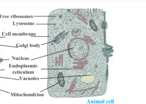
Cell Structure –
- Cell organelle/ Part of the cell – the parts of the cell are as follows –
- 1) Non Living cell organelle –
- a) Cell wall
- 2) Living Cell organelle –
- a) Cell Membrane
- 3) Cell organelle present in Nucleus –
- a) Nucleoplasm
- b) Nuclear Membrane
- c) Nucleoli
- d) Chromatin Fiber
- 4) Cell Organelle Present in Cytoplasm –
- 1) Endoplasmic Reticulum ( ER)
- 2) Golgi apparatus
- 3) Ribosome
- 4) Mitochondria
- 5) Plastid
- 6) Centrosome
- 7) Granules
- 8) Vacuoles
- 9) Fat Droplets
- 10) Lysosome
-

Cell – Unit of life class 9 Selina Biology -
1) Cell wall –
- – it is only present in plant cell
- It is Surrounded to the cell membrane
- cell wall is made up of ” Cellulose”
- It is Freely Permeable
- It gives shape to the cell
-
2) Cell Membrane –
- Cell membrane is made up of a Protein known as ” Lipoprotein”
- It is selectively Permeable
- It is also known as Plasma Membrane
-
3) Cytoplasm –
- State – It is Semi- Liquid in state
- Appearance – It is Colourless
- Many cell organelle are present in the Cytoplasm
- following cell organelle are present in cytoplasm
-
1) Endoplasmic Reticulum (ER) –
- – it is a Network of small/Tiny Tubular Structure Scattered in the Cytoplasm
– it is Classified into 2 Types Based on the Attachment Ribosome
a) Rough Endoplasmic Reticulum
b) Smooth Endoplasmic Reticuluma) Rough Endoplasmic Reticulum –
– the Endoplasmic Reticulum have/ bearing Ribosome on their Surface is Known as Rough Endoplasmic Reticulum
– These are Involved in Protein Synthesis
b) Smooth Endoplasmic Reticulum –
– When Ribosome are not attached with Endoplasmic Reticulum it is known as Smooth Endoplasmic Reticulum
– it is Involved in Synthesis of Lipid - 2) Ribosome –
– It is Discovered by George Palade in 1953
– It is the Site of Synthesis of Protein - 3) Mitochondria –
Shape – It is Cylindrical/ Rod Shaped
– Each Mitochondria is Double Membrane bound Structure with Outer Membrane and Inner Membrane
– The Outer Membrane Forms the Continuous Limiting Boundry of the Organelle
– The Inner Membrane Forms Infolding Known as ” Cristae “
– it is the Place where Respiration Occurs
– Mitochondria are Known as “Power House of the Cell”
– Mitochondria Contain Own DNA - 4) Golgi Apparatus –
– It was 1st Observed By ” Camillo Golgi” In 1898 hence It is later Named after after Him as “Golgi Body ”
– it is Composed of Large Number ” Cisternae”
– it is the Important Site of Formation of “Glycoprotein” and “Glycolipid” - 5) Lysosome –
– Lysosome is also Known as ” Suicide Bag of the Cell “ - 6) Plastids –
– Plastids are only Found in
a) Plants
b) Euglenoids
– They Possess specific Pigment
– on the basis of Pigments Plastids are Classified as Follows -
1) Leucoplast
2) Chromoplast
3) Chloroplast - 1) Leucoplast –
– The Leucoplast are the Colourless Plastids
– They are Varied in different Shapes and different Sizes
– Amyloplast – It Stores Carbohydrates ( Starch )
Eg – Potato - 2) Chromoplast –
– They are Different Coloured Plastids Like – Red, Yellow, Orange - 3) Chloroplast –
– These are Green Coloured Plastids
– These are Present in Plants
– They Take part in the Process of Photosynthesis -

Leaf Chlorophyll - * Vacuoles –
– It is the Membrane Bound Space Found in the Cytoplasm
– These are Larger in Size in the Plants
– These are Smaller in Size in Animals -
Selina biology Class 9 chapter 2
- * Nucleus –
– Nucleus was 1st Described by ” Robert Brown”
– it Contains Genes
– It is Associated with Cell Division
– It is Spherical in Size - NEET PYQ Chapterwise – Animal Kingdom 1
